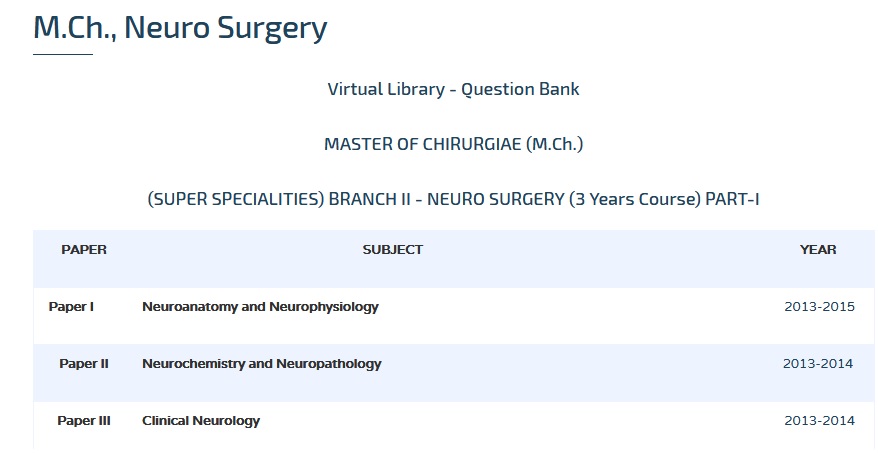
Clinical Neurology & Neuro Radiology M.Ch Question Bank : web.tnmgrmu.ac.in
Name of the University : The Tamilnadu Dr. M.G.R. Medical University
Degree : MASTER OF CHIRURGIAE (M.Ch.)
Branch : BRANCH II – NEURO SURGERY
Subject Code/Name : 1582/CLINICAL NEUROLOGY AND NEURO RADIOLOGY
Paper : II
Document Type : Question Bank
Website : web.tnmgrmu.ac.in
Download Model/Sample Question Paper :
(3 Years Course) PART-I :
1997-2000 : https://www.pdfquestion.in/uploads/web.tnmgrmu.ac.in/4542-1-171582KC.pdf
2001-2010 : https://www.pdfquestion.in/uploads/web.tnmgrmu.ac.in/4542-2-171582KU.pdf
Candidates admitted upto 2005-06 to 2007-08 :
(6 Years Course) PART-II :
2009-2013 : https://www.pdfquestion.in/uploads/web.tnmgrmu.ac.in/4542-3-181582KU.pdf
Clinical Neurology & Neuro Radiology Question Paper
Sub. Code: 1582
M.CH DEGREE EXAMINATIONS
(Higher Specialities)
(Revised Regulations for 3 years course)
Branch II – Neuro Surgery
Paper II : CLINICAL NEUROLOGY AND NEURO RADIOLOGY
Q.P. Code: 171582
Time: Three hours
Maximum: 100 Marks
Related : TNMGR Recent Advances in Surgical Oncology M.Ch Question Bank : www.pdfquestion.in/4593.html
February 2009
ANSWER ALL QUESTIONS :
Draw suitable diagrams wherever necessary. :
I. Essays: 2 x 20 = 40 Marks
1. Describe anatomy of tentorial incisura and clinical features of central and lateral herniation.
2. Trace the optic pathways and different types of visual defect on lesions at various planes.

II. Write short notes on: 10 X 6 = 60 Marks
1. Functional MRI.
2. Cauda/conus syndrome.
3. MR Spectroscopy.
4. Cerebellar signs.
5. Type of Diabetes incipidus and management.
6. Radiological difference between Extradural/Intradural/Intramedullary compression.
7. Neuro cutaneous markers and clinical importance.
8. Eye signs in Brain stem and cervicomedullary compression.
9. Clinical sign of hypertonia in upper and lower limbs.
10. Paediatric coma scales.
August 2009
I. Essays: 2 x 20 = 40 Marks
1. Discuss the anatomy of cerebellum and localization of various clinical syndromes.
2. Imaging of a patient with suspected subarachnoid haemorrhage.
II. Write short notes on: 10 X 6 = 60 Marks
1. Magnetic resonance spectroscopy.
2. Cerebral salt wasting syndrome.
3. Jugular foramen syndrome.
4. Paraplegia in flexion.
5. Evidence based medicine.
6. Brain monitoring in ICU.
7. Primitive reflexes.
8. Transcranial Doppler in neurosurgical practice.
9. Migraine with aura.
10. Brain death.
February 2010
I. Essays: 2 x 20 = 40 Marks
1. Classify epilepsy and describe salient features of each type.
2. Discuss differential diagnosis of recurrent quadriparesis.
II. Write short notes on: 10 X 6 = 60 Marks
1. MR imaging of intracerebral haematoma.
2. Hounsfield scale.
3. Optic Neuritis.
4. Persistant vegetative state.
5. Treatment of status epilepticus.
6. Carpal tunnel syndrome.
7. Cushing’s disease.
8. Functional imaging.
9. Complication of endovascular surgery.
10. Nystagmus.
August 2011
I. Elaborate on :
1. Discuss the differential diagnosis and management of sudden onset right hemiplegia in 20 year old man.
2. Discuss the recent advances in CT and MRI and their applications in Neurological practice.
II. Write notes on :
1. Internuclear ophthalmoplegia.
2. Amaurosis fugax.
3. Lateral medullary syndrome.
4. Medical treatment of migraine.
5. Newer anticonvulsants.
6. Radiological diagnosis of brain abscess.
7. Diagnostic evaluation of Normal pressure hydrocephalus.
8. MRI appearance of intramedullary spinal cord tumours.
9. Ultrasound in Neurosurgery.
10. Diffusion tensor imaging.
August 2012
I. Elaborate on:
1. Discuss the anatomical differential diagnosis of a patient presenting with paraparesis. Mention the clinical findings that would help in arriving at a diagnosis with the anatomical basis for the same.
2. Discuss the differential diagnosis in a 45 year old man brought with a diagnosis of sudden onset inability to speak with weakness of the right upper limb for the past one hour. Mention the relevant radiological investigations that would guide in the immediate management of this patient. Briefly outline the management of this patient during the first twenty four hours of his admission.
II. Write notes on:
1. False localizing signs in clinical neurological examination.
2. Petit mal epilepsy.
3. Approach to a patient with bibrachial amyotrophy.
4. Transcranial Doppler.
5. Moyamoya disease.
6. Radiological findings in tuberculous meningitis and their pathological basis.
7. Sodium Valproate.
8. Differential diagnosis of tremor.
9.Papilloedema.
10. Apraxia.
August 2013
Sub. Code: 1582
I. Elaborate on: (2X15=30)
1. Discuss the differential diagnosis in a patient presenting with decreased hearing in one ear with ipsilateral facial paresis, diminished facial sensations and ataxia. What are the radiological investigations that would aid in arriving at a diagnosis?
2. Discuss the differential diagnosis in a fifty year old man with a ring nhancing lesion in the right frontal lobe, who presented with one episode of generalized seizure. Mention the radiological investigations which would aid in the diagnosis.
II. Write notes on: (10X7=70)
1. Sensory ataxia.
2. Tactile agnosia.
3. Fungal meningitis.
4. Spontaneous intracerebral haemorrhage.
5. Supplementary motor area.
6. Nystagmus.
7. Papilloedema.
8. Chorea.
9. Hemianopia.
10. Brain mass in an HIV seropositive individual.